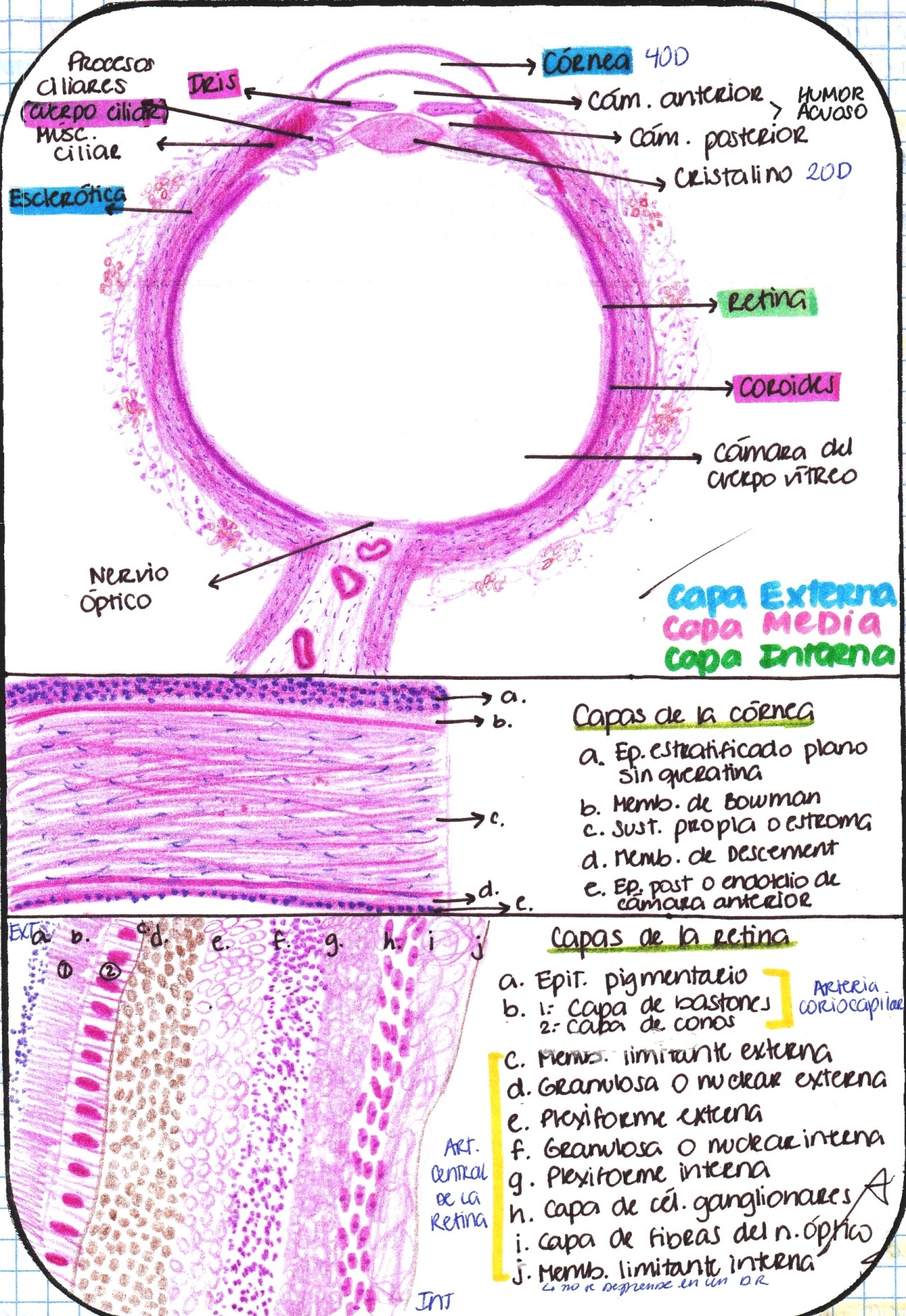

The very central part of the macula is called the fovea. This central part of the retina is called the macula. The centre of the retina is responsible for central vision (looking straight ahead). As it is devoid of rods and cones it forms a physiological “blind spot” within the field of vision. 1.2 million nerve fibres leave the retina at the lamina cribosa of the optic disc to form the optic nerve. The optic nerve consists of ganglion cell axons from the neurosensory retina. At the macula (central retina) the nerve fibre layer may consist of seven layers of ganglion cell bodies whereas in the peripheral retina it may consist of only one. Their axons leave the eye through the lamina cribrosa to form the optic nerve. The cell bodies of the ganglion cells are situated between the nerve fibre layer and the inner plexiform (“synaptic”) layer.

In the peripheral retina one bipolar cell may synapse with up to one hundred rods. At the fovea, one bipolar cell synapses with one photoreceptor and one ganglion cell. The cell bodies of the bipolar cells lie in the inner nuclear layer. Over 35 million bipolar cells connect the photoreceptors to the ganglion cells. The periphery of the retina contains mainly rods (30,000 per mm2) whereas the fovea consists only of cones (150,000 per mm2). The three main neural cell types responsible for relaying the impulses generated by light are: The neurosensory retina is transparent and is thinnest at the fovea. The neurosensory retina, or inner layer, consists of three layers of nuclei (ganglion cell, inner and outer nuclear cell layers) and three layers of fibres (nerve fibre, inner and outer plexiform layers). 90% of the blood that circulates to the eye goes to the choroid, reflecting demands of the photoreceptors and their “supporting” retinal pigment epithelial cells. These photoreceptors are nourished by the retinal pigment epithlelial cells that receive their blood supply from the underlying choroid. The retina is the most metabolically active part of the eye as it contains the photoreceptors. Although the retina is connected to the brain by the optic nerve, the optic nerve behaves more like a white matter tract as it is incapable of regeneration. The retina is the “seeing” part of the eye and is a direct continuation of the brain. This circular “strip”, devoid of retina, is termed the pars plana and is the main site of attachment for the vitreous gel. Unlike the sclera and the choroid, which continue forwards to form the sclera and the iris respectively, the retina stops shortly before the ciliary body. The retina is the inner most layer of the eye.


 0 kommentar(er)
